
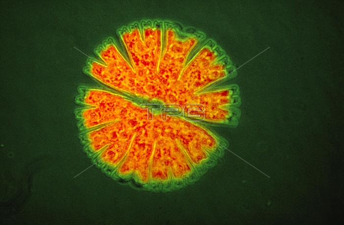
Ultraviolet fluorescence light micrograph of a single- cell desmid Micrasterias sp. The cell is divided into two halves, separated by a narrow waist. Division of the desmid occurs by splitting in two at the waist, with each half generating a perfect replica of itself to restore the original shape. When exposed to ultraviolet light, chlorophyll, the light sensitive pigment contained within green algae, fluoresces intensely. The red coloured areas in this micrograph correspond to chlorophyll in the cell.
| px | px | dpi | = | cm | x | cm | = | MB |
Details
Creative#:
TOP10167483
Source:
達志影像
Authorization Type:
RM
Release Information:
須由TPG 完整授權
Model Release:
N/A
Property Release:
N/A
Right to Privacy:
No
Same folder images:
 Loading