
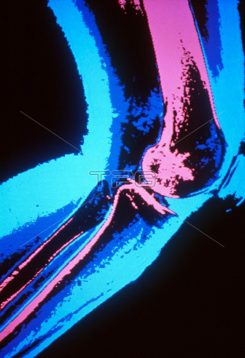
Normal knee. Coloured X-ray of the human knee seen from the side while bent. Four bones meet at the knee, two of which form a joint that works like a hinge. The large bone at the top is the femur, or thigh bone (pink). Below this are the tibia, or shin bone, and its smaller neighbour the fibula (left). At centre right is the patella, or kneecap (blue). The rounded bottom surface of the femur forms a narrow ridge that moves through a groove in the top of the tibia. This arrangement allows to and fro movement only, although slight rotation can occur when the knee is bent.
| px | px | dpi | = | cm | x | cm | = | MB |
Details
Creative#:
TOP10217050
Source:
達志影像
Authorization Type:
RM
Release Information:
須由TPG 完整授權
Model Release:
N/A
Property Release:
N/A
Right to Privacy:
No
Same folder images:
 Loading