
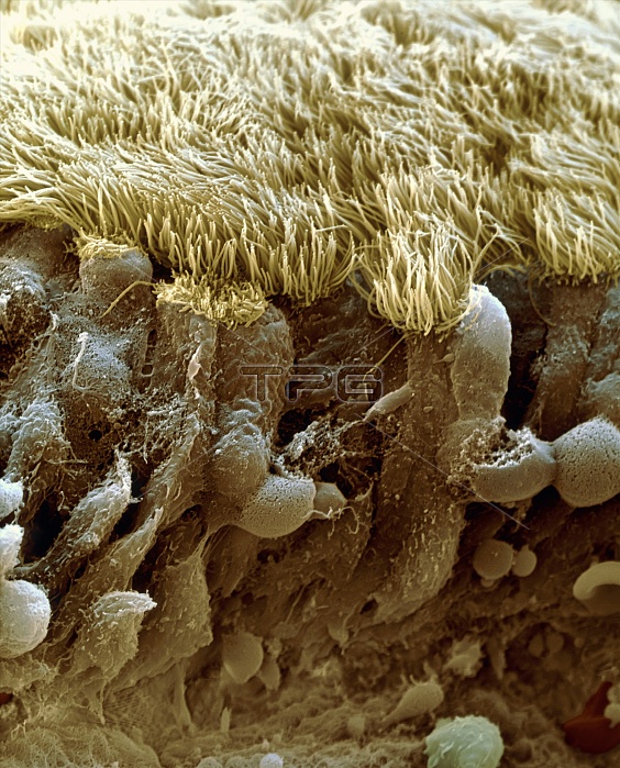
Nasal lining. Coloured scanning electron micrograph (SEM) of a section through the epithelial lining of the nasal cavity. The cylindrical epithelial cells (dark brown, lower frame) are topped with microscopic hair-like structures known as cilia (yellow). These are covered with a sticky mucus (not seen) that traps dust and other inhaled particles. Coordinated, wave-like beating of the cilia propels the mucus to the back of the nose (pharynx), where it is swallowed. Magnification: x1,230 at 6x7cmsize. x2000 at 4x5"~PORTRAIT"
| px | px | dpi | = | cm | x | cm | = | MB |
Details
Creative#:
TOP10219730
Source:
達志影像
Authorization Type:
RM
Release Information:
須由TPG 完整授權
Model Release:
N/A
Property Release:
N/A
Right to Privacy:
No
Same folder images:
 Loading