
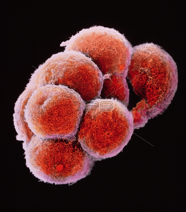
Embryo development. False-colour scanning electron micrograph of an embryo at the early stage known as the morula. The egg reaches this phase about 4 days after fertilisation after a series of mitotic divisions. At this stage about 12-16 cells are present and are surrounded by a thin glycoprotein layer, the zona pellucida, which was here removed. The inner cells of the morula will give rise to the tissues of the embryo while the outer cells, covered here by microvilli (tiny orange ridges), will form the placenta. The morula will implant into the uterus six days after fertilisation. Magnification: x645 at 6x7cm size. Magnification: x1005 at 4x5 inch size. This is a mouse morula.
| px | px | dpi | = | cm | x | cm | = | MB |
Details
Creative#:
TOP10222158
Source:
達志影像
Authorization Type:
RM
Release Information:
須由TPG 完整授權
Model Release:
N/A
Property Release:
N/A
Right to Privacy:
No
Same folder images:
 Loading