
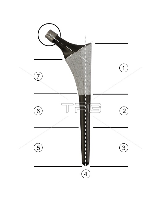
Prosthetic hip joint and Gruen zones. Diagram of the femoral component of a hip prosthesis with labels indicating the hip connection (upper left) and the seven Gruen zones. This component is implanted in the femur (thigh bone) after the head of the femur has been surgically removed. The other components (not shown) are a rounded ball on the insertion point to fit into the socket implanted in the patient's pelvis. Gruen zones are used to assess bone mineral density (BMD) after total hip replacement surgery. This is a Symax prosthesis. For a set of Gruen zone diagrams, see C016/6776 to C016/6781.
| px | px | dpi | = | cm | x | cm | = | MB |
Details
Creative#:
TOP11716555
Source:
達志影像
Authorization Type:
RM
Release Information:
須由TPG 完整授權
Model Release:
NO
Property Release:
NO
Right to Privacy:
No
Same folder images:
 Loading