
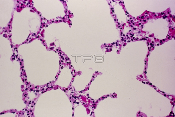
Lung alveoli. Light micrograph of a cross section through lung tissue showing alveoli. Making up the frame (spongy appearance) are tiny air sacs called alveoli. The alveolar walls (pink) are lined with a one-cell thick layer of epithelium. Breathed in air diffuses through the epithelium to a rich network of capillaries in the walls. This is where exchange of gases (oxygen and carbon dioxide) takes place. Oxygen will be carried by the capillary blood to the heart. At the same time carbon dioxide will diffuse out of the blood into the lung and will be breathed out. Haematoxylin and eosin stained. Magnification unknown.
| px | px | dpi | = | cm | x | cm | = | MB |
Details
Creative#:
TOP11722288
Source:
達志影像
Authorization Type:
RM
Release Information:
須由TPG 完整授權
Model Release:
NO
Property Release:
NO
Right to Privacy:
No
Same folder images:
 Loading