
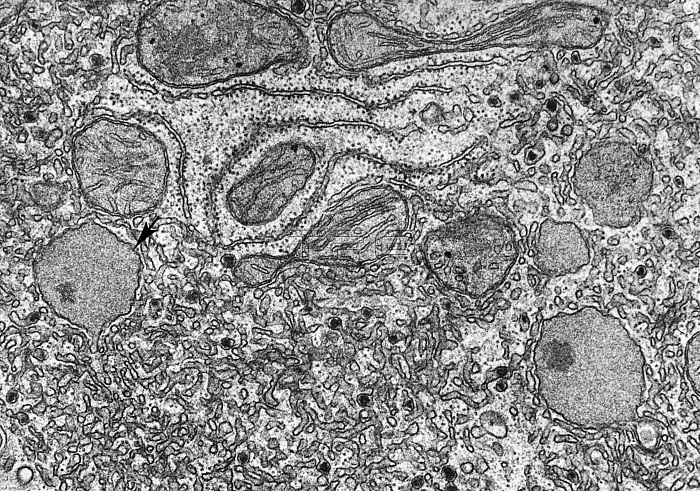
Transmission electron micrograph of an area of cytoplasm from a rat liver cell containing peroxisomes. Peroxisomes are abundant in hepatic cells and were first isolated from the crude lysosome fraction of liver homogenates. Visible at the arrows, peroxisomes are generally spherical and have homogeneous content of relatively low density.
| px | px | dpi | = | cm | x | cm | = | MB |
Details
Creative#:
TOP22218881
Source:
達志影像
Authorization Type:
RM
Release Information:
須由TPG 完整授權
Model Release:
N/A
Property Release:
No
Right to Privacy:
No
Same folder images:
 Loading