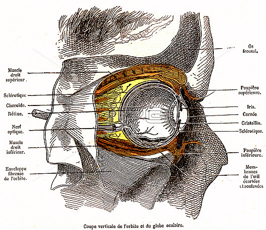
Eye anatomy, 19th-century illustration. Sagittal section of the orbit of the eye and the eye ball. The labels include structures such as the frontal bone, the iris, the cornea, the lens, the sclera, the optic nerve, various muscles, the retina, and the choroid. The labels are in French. Published in 'La Vie Normale et la Sante' (Normal Life and Health, Paris, 1881) by Dr Jules Rengade.
| px | px | dpi | = | cm | x | cm | = | MB |
Details
Creative#:
TOP26513879
Source:
達志影像
Authorization Type:
RM
Release Information:
須由TPG 完整授權
Model Release:
N/A
Property Release:
N/A
Right to Privacy:
No
Same folder images:
1800sanatomicalbiologicalchoroidcorneaeuropeaneyeballeyesocketeyeballfrenchfrenchlanguagefrontalbonehealthyhistoricalirisjulesrengadelavienormaleetlasantelabellabeledlabelledlabelslaterallensmusclesno-onenobodynormalnormallifeandhealthocularophthalmicopticnerveorbitoftheeyeretinasagittalsclerasectionsectionedsideviewtexteyeorganhumanbodyheadanatomybiologyhistoryophthalmologyartworkillustration19thcentury1881
1800s188119thanatomicalanatomyandartworkballbiologicalbiologybodybonecenturychoroidcorneaeteuropeaneyeeyeeyeeyeeyeballfrenchfrenchfrontalheadhealthhealthyhistoricalhistoryhumanillustrationirisjuleslalalabellabeledlabelledlabelslanguagelaterallenslifemusclesnerveno-onenobodynormalnormalnormaleocularofophthalmicophthalmologyopticorbitorganrengaderetinasagittalsantesclerasectionsectionedsidesockettextthevieview

 Loading