
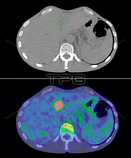
Secondary cancer in the liver. Comparison of axial computed tomography (CT, top) and positron emission tomography (PET, bottom) scans of a patient with kidney cancer that has spread to the liver. The secondary cancer in the liver is not seen on the CT scan, but has been detected by the PET scan as the circular pink area. The primary cancer is of the right kidney, and it has spread (metastasised) to the liver. The PET scan has been obtained with 2-18F-fluoro-2-deoxy-D-glucose (FDG), a radioactive tracer that can be used to identify cancer sites.
| px | px | dpi | = | cm | x | cm | = | MB |
Details
Creative#:
TOP30173517
Source:
達志影像
Authorization Type:
RM
Release Information:
須由TPG 完整授權
Model Release:
N/A
Property Release:
N/A
Right to Privacy:
No
Same folder images:
2-18f-fluoro-2-deoxy-d-glucoseabnormalcancerouscombinedcomputedtomographyconditiondiagnosisdiagnosticdisorderfalse-colouredfdgfluorineglucosehepaticmalignancymalignantmedicalphysicsmetastasismetastaticnuclearmedicinephysicalpositronemissiontomographyradiationradioactiveradioactivetracerscanssecondaryspreadtumourunhealthyaxialsectionsectionedrenalno-onenobodypairduokidneycancerlivercancerdiseasecancerhumanbodyabdomenliverkidneymedicineoncologypatientctscancolouredpetscanscanner2two
22-18f-fluoro-2-deoxy-d-glucoseabdomenabnormalaxialbodycancercancercancercancerouscolouredcombinedcomputedconditionctdiagnosisdiagnosticdiseasedisorderduoemissionfalse-colouredfdgfluorineglucosehepatichumankidneykidneyliverlivermalignancymalignantmedicalmedicinemedicinemetastasismetastaticno-onenobodynuclearoncologypairpatientpetphysicalphysicspositronradiationradioactiveradioactiverenalscanscanscannerscanssecondarysectionsectionedspreadtomographytomographytracertumourtwounhealthy
 Loading