
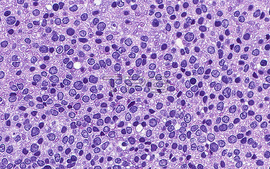
Light micrograph of tubular adenoma cell nuclei in cross section. Tubular adenomas are colon lesions that are precancerous and that are often detected as colon polyps during endoscopic screening for colon cancer. This image shows the nuclei of tubular adenoma cells (round light to dark blue dots) in cross section. Haematoxylin and eosin stained tissue section. Magnification: x600 when printed at 10cm.
| px | px | dpi | = | cm | x | cm | = | MB |
Details
Creative#:
TOP30292966
Source:
達志影像
Authorization Type:
RM
Release Information:
須由TPG 完整授權
Model Release:
N/A
Property Release:
N/A
Right to Privacy:
No
Same folder images:
pathologymedicineanatomicpathologysurgicalpathologycolontubularadenomacolonpolypcrosssectiondysplasiaprecancercancerriskcoloncancerscreeningendoscopygastrointestinalgastrointestinalpathologydigestivesystemgastroenterologyintestineboweltumortumouroncologymasshematoxylinhaematoxylineosinstaindiseasedisorderconditionabnormalunhealthybiologybiologicalhistologyhistologicalhistopathologylightmicrographlmmicroscopynobodyno-oneslidehumanbodyanatomyanatomicaltissuecells
abnormaladenomaanatomicanatomicalanatomybiologicalbiologybodybowelcancercancercellscoloncoloncolonconditioncrossdigestivediseasedisorderdysplasiaendoscopyeosingastroenterologygastrointestinalgastrointestinalhaematoxylinhematoxylinhistologicalhistologyhistopathologyhumanintestinelightlmmassmedicinemicrographmicroscopyno-onenobodyoncologypathologypathologypathologypathologypolypprecancerriskscreeningsectionslidestainsurgicalsystemtissuetubulartumortumourunhealthy
 Loading