
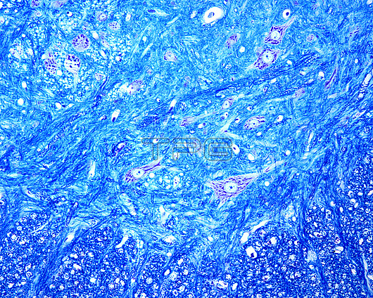
Light micrograph of the ventral horn of a cross-sectioned spinal cord stained with Luxol fast blue stain. The Nissl bodies of motor neurons (nerve cells) are stained dark violet, while myelinated fibres are stained blue, especially in the white matter.
| px | px | dpi | = | cm | x | cm | = | MB |
Details
Creative#:
TOP29820930
Source:
達志影像
Authorization Type:
RM
Release Information:
須由TPG 完整授權
Model Release:
N/A
Property Release:
N/A
Right to Privacy:
No
Same folder images:
spinalcordmyelinatedfiberfibregreymatterhornventralhornnisslbodymotorneuronluxolmicroscopymyelinatednervousspinalsystemwhitematterneurologyneurohistologybiologyhistologyhistologicallightmicrographfiberfibreneurologicalbiologicallightmicrographlmbiologybiologicalhistologyhistologicalnormalhealthynobodyno-one
biologicalbiologicalbiologybiologybodycordfiberfiberfibrefibregreyhealthyhistologicalhistologicalhistologyhistologyhornhornlightlightlmluxolmattermattermicrographmicrographmicroscopymotormyelinatedmyelinatednervousneurohistologyneurologicalneurologyneuronnisslno-onenobodynormalspinalspinalsystemventralwhite
 Loading