
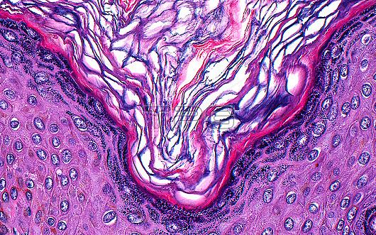
Light micrograph of epidermis. The epidermis is the outer layer of skin. It consists of squamous cells (light pink with blue ovals) and an outermost layer of keratin (pink and purple intersecting lines from centre of image to upper corners). The outermost layer of squamous cells, beneath the keratin layer forms a granular layer (dark purple layer with many small dots). The epidermis projects periodically into the dermis, forming what are known as rete ridges. This is the reason the layers of the epidermis appear higher at the left and right edges of this image ??the rete ridge projects downwards along the central vertical axis of this image. Haematoxylin and eosin stained tissue section. Magnification: x400 when printed at 10cm.
| px | px | dpi | = | cm | x | cm | = | MB |
Details
Creative#:
TOP30292998
Source:
達志影像
Authorization Type:
RM
Release Information:
須由TPG 完整授權
Model Release:
N/A
Property Release:
N/A
Right to Privacy:
No
Same folder images:
 Loading