
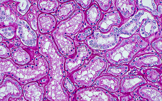
Light micrograph of proximal renal tubules. Proximal real tubules have large amounts of cytoplasm (light pink) and a tortuous course. This image shows cross sections of proximal tubules, outlined by their basement membranes (dark pink lines). The nuclei of the tubules are the small round blue dots. Periodic Acid Schiff (PAS) stained tissue section. Magnification: x200 when printed at 10cm.
| px | px | dpi | = | cm | x | cm | = | MB |
Details
Creative#:
TOP30293016
Source:
達志影像
Authorization Type:
RM
Release Information:
須由TPG 完整授權
Model Release:
N/A
Property Release:
N/A
Right to Privacy:
No
Same folder images:
pathologymedicineanatomicpathologysurgicalpathologykidneyrenaltubulesproximaltubulesrenalpathologymedicalrenalpathologynephrologynormalhistologyurinarytracturinarysystemgenitourinarygenitourinarypathologyurologypasstainbiologybiologicalhistologyhistologicalhistopathologylightmicrographlmmicroscopynobodyno-oneslidehumanbodyanatomyanatomicaltissuecells
anatomicanatomicalanatomybiologicalbiologybodycellsgenitourinarygenitourinaryhistologicalhistologyhistologyhistopathologyhumankidneylightlmmedicalmedicinemicrographmicroscopynephrologyno-onenobodynormalpaspathologypathologypathologypathologypathologypathologyproximalrenalrenalrenalslidestainsurgicalsystemtissuetracttubulestubulesurinaryurinaryurology
 Loading