
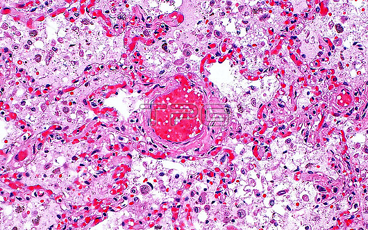
Light micrograph of pulmonary congestion. Pulmonary congestion occurs when blood flow in the lungs is backed up and blood accumulates in vessels. A larger vessel is filled with red blood cells (dark red, centre of image) and smaller pulmonary capillaries are also packed with many red blood cells (bright red, smaller dots and streaks). The pink areas in between vessels represent the septa (lining structures) of the lung alveolar space (white). The alveolar spaces also contain oedema (light pink) and some macrophages (slightly darker pink dots). Haematoxylin and eosin stained tissue section. Magnification: x200 when printed at 10cm.
| px | px | dpi | = | cm | x | cm | = | MB |
Details
Creative#:
TOP30293018
Source:
達志影像
Authorization Type:
RM
Release Information:
須由TPG 完整授權
Model Release:
N/A
Property Release:
N/A
Right to Privacy:
No
Same folder images:
 Loading