
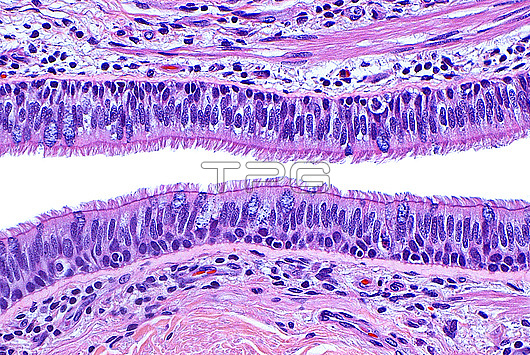
Light micrograph of respiratory epithelium. The epithelium (surface layer of cells) lining areas of the respiratory tract is composed of cells with columnar or elongated nuclei (dark blue-purple) and cilia (many thin hair-like pink lines). Occasional goblet cells (larger and more blue ovoid structures) are scattered in the epithelium, and these goblet cells secrete mucin (mucous). Beneath the epithelium is connective tissue (pink at top and bottom of image) which contains blood vessels (dark red). Haematoxylin and eosin stained tissue section. Magnification: x200 when printed at 10cm.
| px | px | dpi | = | cm | x | cm | = | MB |
Details
Creative#:
TOP30293027
Source:
達志影像
Authorization Type:
RM
Release Information:
須由TPG 完整授權
Model Release:
N/A
Property Release:
N/A
Right to Privacy:
No
Same folder images:
pathologymedicineanatomicpathologysurgicalpathologyrespiratoryepitheliumrespiratorysystemciliacolumnarcellsgobletcellsmucousmucinpulmonarypulmonologyenthematoxylinhaematoxylineosinstainnormalhistologybenignbiologybiologicalhistologyhistologicalhistopathologylightmicrographlmmicroscopynobodyno-oneslidehumanbodyanatomyanatomicaltissuecells
anatomicanatomicalanatomybenignbiologicalbiologybodycellscellscellsciliacolumnarenteosinepitheliumgoblethaematoxylinhematoxylinhistologicalhistologyhistologyhistopathologyhumanlightlmmedicinemicrographmicroscopymucinmucousno-onenobodynormalpathologypathologypathologypulmonarypulmonologyrespiratoryrespiratoryslidestainsurgicalsystemtissue
 Loading