
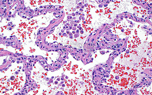
Light micrograph of lung with septal fibrosis. Fibrosis in the lungs can occur following chronic injury. One form of fibrosis leads to widening of the alveolar septa (pink curved lines). The alveoli also contain histiocytes (round pink dots), red blood cells (dark red dots) and oedema fluid (very light blue within the white spaces). Haematoxylin and eosin stained tissue section. Magnification: x200 when printed at 10cm.
| px | px | dpi | = | cm | x | cm | = | MB |
Details
Creative#:
TOP30293031
Source:
達志影像
Authorization Type:
RM
Release Information:
須由TPG 完整授權
Model Release:
N/A
Property Release:
N/A
Right to Privacy:
No
Same folder images:
pathologymedicineanatomicpathologysurgicalpathologylungpulmonarypulmonarypathologyautopsyfibrosisthoracicpathologypulmonologyrespiratorysystemrespiratorydiseasediseasedisorderconditionabnormalunhealthyhematoxylinandeosinbiologybiologicalhistologyhistologicalhistopathologylightmicrographlmmicroscopynobodyno-oneslidehumanbodyanatomyanatomicaltissuecells
abnormalanatomicanatomicalanatomyandautopsybiologicalbiologybodycellsconditiondiseasediseasedisordereosinfibrosishematoxylinhistologicalhistologyhistopathologyhumanlightlmlungmedicinemicrographmicroscopyno-onenobodypathologypathologypathologypathologypathologypulmonarypulmonarypulmonologyrespiratoryrespiratoryslidesurgicalsystemthoracictissueunhealthy
 Loading