
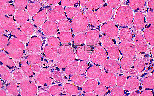
Light micrograph of skeletal muscle cells. The skeletal muscle cells have a large cytoplasm (pink) which contains the contractile fibres (very thin lines). There are several nuclei (dark blue dots) per cell, and the nuclei are arranged at the periphery of the cells. Haematoxylin and eosin stained tissue section. Magnification: x400 when printed at 10cm.
| px | px | dpi | = | cm | x | cm | = | MB |
Details
Creative#:
TOP30293040
Source:
達志影像
Authorization Type:
RM
Release Information:
須由TPG 完整授權
Model Release:
N/A
Property Release:
N/A
Right to Privacy:
No
Same folder images:
pathologymedicineanatomicpathologysurgicalpathologyskeletalmusclemusclesofttissuemusculoskeletalsystemsofttissuepathologynormalhistologybenignorthopedicsorthopaedicsorthopedicorthopaedicboneandsofttissuepathologyhematoxylinandeosinbiologybiologicalhistologyhistologicalhistopathologylightmicrographlmmicroscopynobodyno-oneslidehumanbodyanatomyanatomicaltissuecells
anatomicanatomicalanatomyandandbenignbiologicalbiologybodybonecellseosinhematoxylinhistologicalhistologyhistologyhistopathologyhumanlightlmmedicinemicrographmicroscopymusclemusclemusculoskeletalno-onenobodynormalorthopaedicorthopaedicsorthopedicorthopedicspathologypathologypathologypathologypathologyskeletalslidesoftsoftsoftsurgicalsystemtissuetissuetissuetissue
 Loading