
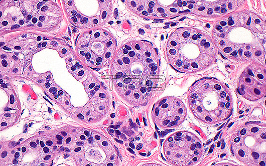
Light micrograph of sweat (eccrine) glands. The sweat glands are found in the connective tissue of the skin dermis (pink). They secrete sweat into their lumens (white) which is excreted at the surface of skin. The sweat gland cells have eosinophilic cytoplasm (darker pink) and round nuclei (blue dots), and are arranged circumferentially around the lumens. Haematoxylin and eosin stained tissue section. Magnification: x200 when printed at 10cm.
| px | px | dpi | = | cm | x | cm | = | MB |
Details
Creative#:
TOP30293046
Source:
達志影像
Authorization Type:
RM
Release Information:
須由TPG 完整授權
Model Release:
N/A
Property Release:
N/A
Right to Privacy:
No
Same folder images:
pathologymedicineanatomicpathologysurgicalpathologysweatglandssweateccrineskindermatologydermatologynormalhistologyintegumentarysystemglandexocrineglandhematoxylinandeosinbiologybiologicalhistologyhistologicalhistopathologylightmicrographlmmicroscopynobodyno-oneslidehumanbodyanatomyanatomicaltissuecells
anatomicanatomicalanatomyandbiologicalbiologybodycellsdermatologydermatologyeccrineeosinexocrineglandglandglandshematoxylinhistologicalhistologyhistologyhistopathologyhumanintegumentarylightlmmedicinemicrographmicroscopyno-onenobodynormalpathologypathologypathologyskinslidesurgicalsweatsweatsystemtissue
 Loading